

Can A New Ex-Fix Device Have An Impact In Deformity Correction?
Complications related to obesity have been a topic of concern for health officials since the 1950s. However, it has only been in the past few years that a widespread epidemic has reached alarming proportions in the United States and worldwide, leading to substantial health and economic costs. In the U.S., obesity is largely linked to an increase in the incidence of type II diabetes mellitus and metabolic syndrome. In fact, a recently published study by the Centers for Disease Control and Prevention (CDC) warns that “one-third of Americans born in 2000 could develop diabetes.”1 Accordingly, managing associated complications from this disease is an essential priority for those of us who see patients with diabetes on a regular basis. There is an array of lower extremity complications secondary to diabetes. These complications include peripheral vascular disease, peripheral neuropathy and biomechanical deformities resulting from motor-sensory neuropathy. The most notable deformities arise from Charcot neuroarthropathy. Charcot destruction does not occur in the absence of peripheral neuropathy and approximately 30 to 50 percent of patients with diabetes have peripheral neuropathy.2-6 Unfortunately, there is a significant discrepancy in the literature when it comes to the incidence of Charcot in patients with diabetes as the incidence ranges from 0.2 to 29 percent.2,5-10 While Charcot represents one end of the spectrum of diabetic foot deformities, longstanding, motorsensory neuropathy secondary to diabetes represents the other end of the spectrum. On average, a majority of the mild to moderate foot deformities are amenable to accommodative shoe gear, conservative management and/or bracing. Historically, surgery was indicated in a diabetic patient only if he or she had a deep space infection with or without osteomyelitis, when accommodative methods failed or at the patient’s request.4,11
A Closer Look At Surgery For Lower-Extremity Diabetic Deformities
However, the concept of surgical correction for the diabetic foot and ankle deformity has resurfaced in the past few years. Pinzur collected data over a 10-year period on treating Charcot arthropathy of the foot and ankle. According to the data, 48.5 percent of patients were treated with conservative therapy while 50.6 percent of patients underwent surgery. The surgical procedures included 21 major limb amputations, 29 ankle fusions, 26 hindfoot fusions, 23 exostectomies and 23 debridements for osteomyelitis.4 Conservative management of Charcot deformities (stage I or stage II), in the form of total contact casts, bracing devices and/or accommodative footgear, is approximately 75 percent effective.5,12 Surgical reconstruction of a moderate to severe foot and/or ankle deformity secondary to diabetes has come to the limelight as a potential salvage procedure for preventing further deformity and subsequent tissue breakdown, infection and ultimately amputation.9,11,13-16 Myerson and Edwards found that surgical correction resulted in lower-extremity stabilization in 93 percent of patients who presented with severe deformity.5
Assessing The Potential Of The EZ Frame
With this in mind, let us consider a new external fixation device that is specifically used in foot correction and eliminates the need for invasive fixation devices above the ankle. The device is called the EZ Frame (Signal Medical Corp.). It is composed of a boot with adjustable straps, a rocker-bottom footplate, stainless steel posts, wire nuts, smooth wires and olive wires. The boot is connected to the footplate via telescoping carbon fiber rods. Additionally, one can remove the full-length tongue during dressing changes and adjust the positions of both the boot and the frame. Various sizes are available. The footplate also allows versatility in offloading ulcerated areas. In a study performed on seven cadaver specimens and six polyurethane foam models, researchers created a midtarsal wedge osteotomy with a template. They placed an F-Scan pressure insole system (Tekscan) inside the osteotomy and used it to measure and record the pressure created across the osteotomy site after applying fixation. Researchers compared multiple ring external fixation with tension and compression versus the EZ Frame with tension and compression. The F-Scan results demonstrated superior compression strength across the osteotomy fixated with the EZ Frame.17 Researchers believe this superior compression results from the force vectors that are stabilized against the plantar plate apparatus during tensioning of the EZ Frame. The EZ Frame, which was recently FDA-approved, also allows guarded weightbearing. There is a decreased learning curve for this frame as opposed to other external fixation devices. It is also available as a complete system, which reduces OR time. Some of the indications for the EZ Frame include isolated rearfoot arthrodesis, midfoot arthrodesis/osteotomy, comminuted trauma and diabetic Charcot reconstruction. One may also employ this device for the majority of foot pathology that does not require fixation above the ankle.
Case Study: Treating A Patient With Severe PTTD
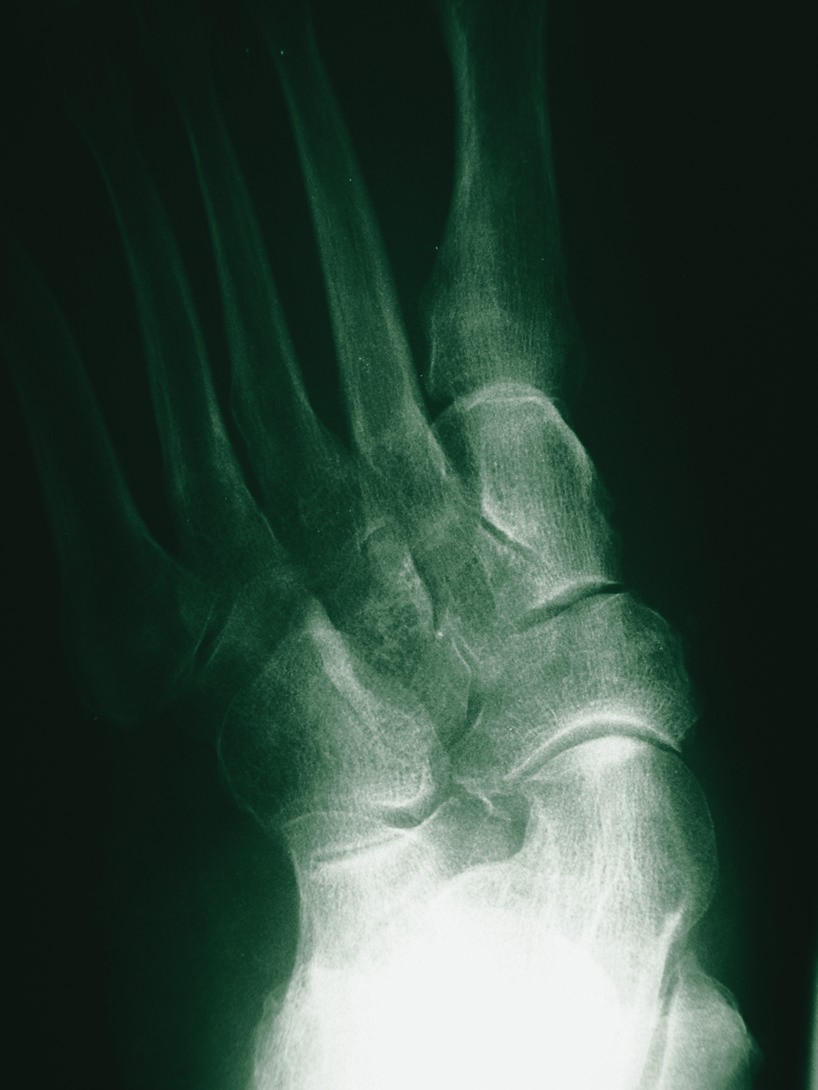
A diabetic patient presented with severe posterior tibial tendon dysfunction and pes plano valgus deformity. Surgeons performed a triple arthrodesis and applied the EZ Frame with four pins. The pins were located in the calcaneus, talus, navicular and across the metatarsals (see right photo). The patient should remain non-weightbearing for up to six weeks. At this time, practitioners can remove the frame and place the patient in a below knee walking cast for approximately two weeks following serial radiographs. One can subsequently transition the patient into a CAM-Walker and eventually into regular shoe gear. The obvious advantages to employing this modality for diabetic foot reconstruction are guarded weightbearing, enhanced stability and the preclusion of proximal pin sites. Dr. Duckworth is a first-year resident at the Southeast Michigan Surgical Hospital in Warren, Mich. Dr. Steinberg (pictured) is an Assistant Professor in the Department of Surgery at the Georgetown University School of Medicine in Washington, D.C. He is a Fellow of the American College of Foot and Ankle Surgeons.
References:
1. www.diabetes.about.com
2. Garapati R, Weinfeld SB. Complex Reconstruction of the Diabetic Foot and Ankle. Am J Surg. 2004; 187(supp): 81s-86s.
3. Glycemic Control and Complications in Non-Insulin Dependent Diabetes Mellitus (A Feasibility Study): VA Cooperative Study No. 344. Washington, D.C., Department of Veterans Affairs.
4. Pinzur MS. Benchmark Analysis of Diabetic Patients with Neuropathic (Charcot) Foot Deformity. Foot & Ankle Int. 1999; 20(9): 564-567.
5. Myerson MS, Edwards WH. Management of Neuropathic Fractures in the Foot and Ankle. J Am Acad Orthop Surg. 1999; 7: 8-18.
6. Schon LC, Easley ME, Weinfeld SB. Charcot Neuroarthropathy of the Foot and Ankle. Clin Orthop & Resch. 1998; 349: 116-131.
7. Herbst SA, Jones KB, Saltzman CL. Pattern of Diabetic Neuropathic Arthropathy Associated with Peripheral Bone Mineral Density. J Bone Joint Surg [Am]. 2004; 86-B(3): 378-383.
8. Fabrin J, Larsen K, Holstein PE. Incidence of Charcot: Long-Term Follow-up in Diabetic Charcot Foot with Spontaneous Onset. 2000; 23(6): 796-800.
9. Jolly GP, Zgonis T, Polyzois V. External Fixation in the Management of Charcot Neuroarthropathy. Clin Podiatr Med Surg. 2003; 20: 741-756.
10. Eichenholtz SN. Charcot Joints. Springfield (IL): C.C. Thomas, 1966.
11. Schon LC, Marks RM. The Management of Neuropathic Fracture-Dislocations in the Diabetic Patient. Orthop Clin N Am. 1995; 26(2): 375-392.
12. Myerson M, Papa J, Eaton K, Wilson K. The Total-Contact Cast for Management of Neuropathic Plantar Ulceration of the Foot. J Bone Joint Surg [Am]. 1992; 74-A(2): 261-269.
13. Pinzur MS, Shields N, Trepman E, Dawson P, et. al. Current Practice Patterns in the Treatment of Charcot Foot. Foot & Ankle Int. 2000; 21(11): 916-920
14. Cooper PS. Application of External Fixators for Management of Charcot Deformities of the Foot and Ankle. Foot Ankle Clin N Am. 2002; 7: 207-254.
15. Farber DC, Juliano PJ, Cavanagh PR, Ulbrecht J, et. al. Single Stage Correction with External Fixation of the Ulcerated Foot in Individuals with Charcot Neuroarthropathy. Foot & Ankle Int. 2002; 23(2): 130-134.
16. Prokuski LJ, Saltzman CL. External Fixation for the Treatment of Charcot Arthropathy of the Ankle: A Case Report. Foot & Ankle Int. 1998; 19(5): 336-341.
17. Grant WP, Rubin L, Pupp G, Vito G, et. al. Mechanical Testing of Seven Fixation Methods For Generation of Compression Across a Midtarsal Osteotomy: A Comparison of Internal and External fixation Devices. In publication.


